What Is Srs Surgery A Precision Revolution In Brain Treatment
Published
Stereotactic radiosurgery SRS represents a groundbreaking fusion of medical innovation and surgical precision reshaping modern neurosurgery forever Unlike traditional open-brain procedures SRS delivers concentrated radiation beams to treat abnormalities with unparalleled accuracy without requiring invasive incisions This non-invasive approach has transformed the management of brain tumors vascular malformations and functional disorders offering patients a less risky alternative to conventional treatments
The journey of SRS began over seven decades ago when early pioneers experimented with focused radiation to target deep-seated brain lesions Today advanced technologies like Gamma Knife CyberKnife and LINAC-based systems enable sub-millimeter precision delivering high-dose radiation directly to tumors while sparing surrounding healthy tissue What makes SRS truly revolutionary is its ability to combine cutting-edge imaging robotics and radiation therapy into a single outpatient procedure capable of addressing conditions once deemed inoperable
Understanding Stereotactic Radiosurgery (SRS): Foundations and Evolution
Stereotactic Radiosurgery (SRS) represents a paradigm shift in neurosurgical treatment, merging precision engineering with advanced radiotherapy to target brain abnormalities without invasive procedures. Unlike traditional open-brain surgery, SRS delivers highly focused radiation beams to tumors, vascular malformations, or functional disorders while sparing surrounding healthy tissue. Its development reflects a convergence of neuroscience, medical physics, and robotics, transforming the landscape of minimally invasive therapies. This section explores the foundational principles of SRS, its distinction from conventional radiotherapy, and the technological milestones that propelled it into modern clinical practice.
Core Principles of Stereotactic Radiosurgery
SRS operates on three foundational pillars: sub-millimeter precision targeting, conformal radiation delivery, and minimal invasiveness. The technique leverages 3D imaging modalities such as MRI and CT scans to create a high-resolution map of the brain, identifying the exact coordinates of the treatment target. Robotics or specialized frames then immobilize the patient’s head, ensuring the target remains stationary during radiation delivery. Unlike conventional radiotherapy, which spreads radiation over multiple sessions to cover larger areas, SRS concentrates dose intensity into a single or few sessions, maximizing tumor control while reducing collateral damage.
The precision of SRS is achieved through stereotactic coordination systems, which integrate linear accelerators (LINACs), cobalt-60 sources (Gamma Knife), or robotic arms (CyberKnife) to deliver radiation from multiple angles. This multi-directional approach creates a tightly conformed dose distribution, resembling a "crossfire" that converges on the target while rapidly falling off outside it. The result is a high dose to the lesion with minimal exposure to adjacent critical structures, such as the optic nerves, brainstem, or motor cortex.
Historical Development of SRS: Milestones and Innovators
The evolution of SRS traces back to the early 20th century, when neurosurgeons sought non-invasive alternatives to treat brain pathologies. Key milestones include:
1949: Introduction of Stereotactic Frames
The first stereotactic frame, developed by Lars Leksell in Sweden, enabled precise localization of brain targets using X-rays. This innovation laid the groundwork for stereotactic neurosurgery, though initial applications focused on biopsy and lesioning rather than radiation.
1951: First Gamma Knife Prototype
Leksell collaborated with Börje Larsson to design the Gamma Knife, initially using a single cobalt-60 source. The device was later refined into a 201-source array in 1968, allowing for more conformal dose distributions.
The linear accelerator (LINAC) was adapted for SRS by John Adler and colleagues at Stanford University. This approach offered greater flexibility in beam shaping and dose planning compared to fixed Gamma Knife units.
1994: FDA Approval of Gamma Knife
The Gamma Knife 2000 received FDA approval, marking the first dedicated SRS device to gain regulatory clearance in the U.S. This milestone accelerated adoption in clinical practice.
1999: CyberKnife System Introduced
Developed by John R. Adler Jr., the CyberKnife combined image-guided robotics with real-time tracking, enabling treatment of moving targets (e.g., tumors near respiratory structures) without rigid immobilization.
2010s: Advancements in MRI-Guided SRS
Integration of intraoperative MRI (e.g., BrainLab’s ExacTrac) and adaptive planning further refined SRS, allowing dynamic adjustments during treatment to account for anatomical changes.
Comparison of SRS to Conventional Radiotherapy
While both SRS and fractionated radiotherapy use ionizing radiation to treat brain tumors, their delivery methods, precision, and clinical applications differ significantly. The following table contrasts the two approaches:
Feature
Stereotactic Radiosurgery (SRS)
Conventional Radiotherapy
Delivery Method
Single or few high-dose sessions (e.g., 1–5 fractions). Uses LINAC, Gamma Knife, or CyberKnife.
Multiple low-dose fractions (e.g., 15–30 sessions) over weeks. Typically delivered via LINAC or proton therapy.
Precision
Sub-millimeter accuracy (≤1 mm margin). Targets confined to small volumes (e.g., ≤4 cm diameter).
Millimeter-level accuracy (3–5 mm margin). Used for larger or diffuse tumors.
Typical Use Cases
Small brain metastases (≤3 cm).
Arteriovenous malformations (AVMs).
Trigeminal neuralgia.
Functional disorders (e.g., essential tremor).
Large or infiltrative tumors (e.g., glioblastoma).
Requires advanced imaging (MRI/CT) and specialized equipment.
Relies on standard LINACs or proton therapy centers.
The high-dose, single-fraction approach of SRS makes it ideal for small, well-defined targets, while conventional radiotherapy’s fractionated dosing is better suited for larger or radiosensitive tissues. However, advancements in hypofractionated SRS (e.g., 3–5 fractions) are bridging this gap for intermediate-sized tumors.
Evolution of SRS Techniques: A Timeline of Technological Advancements
The progression of SRS technology reflects innovations in imaging, robotics, and radiation physics. The following table outlines key developments from early stereotactic frames to contemporary systems:
Year
Milestone
Contributors/Institutions
Impact on Clinical Practice
1949
First stereotactic frame for brain biopsy.
Lars Leksell (Karolinska Institute, Sweden).
Enabled precise targeting of deep brain structures.
1951
Prototype Gamma Knife with single cobalt-60 source.
Leksell & Börje Larsson.
Foundation for non-invasive radiosurgery.
1968
Gamma Knife Model B (201-source array).
Elekta AB (Sweden).
Improved dose conformity for AVMs and tumors.
1980s
LINAC-based SRS development.
John Adler (Stanford University).
Flexibility in beam shaping and dose planning.
1994
FDA approval of Gamma Knife 2000.
<
Medical Conditions Treated with Stereotactic Radiosurgery (SRS)
Stereotactic radiosurgery (SRS) has revolutionized the treatment of various neurological and oncological conditions by delivering precise, high-dose radiation to targeted areas while minimizing damage to surrounding tissues. Unlike conventional radiotherapy, SRS focuses on localized lesions with millimeter accuracy, making it particularly effective for conditions where surgical intervention poses high risks or is impractical. The following sections categorize primary medical conditions addressed by SRS, supported by case studies, mechanistic insights, comparative treatment efficacy, and emerging applications. Data is sourced from peer-reviewed studies, clinical guidelines from the American Society for Radiation Oncology (ASTRO), and global health reports from the National Cancer Institute (NCI).
Categorization of Conditions by Severity and Frequency
Conditions treated with SRS are stratified based on severity (life-threatening vs. chronic/debilitating) and frequency (common vs. rare). This classification aids clinicians in prioritizing treatment protocols and resource allocation. Below are the primary categories:
High Severity and High Frequency
Tumors with high mortality or morbidity rates that frequently require intervention, such as brain metastases (most common indication for SRS) and glioblastoma multiforme (GBM). These conditions demand immediate and effective treatment due to their aggressive nature and limited therapeutic alternatives.
High Severity and Low Frequency
Rare but critical conditions like arteriovenous malformations (AVMs) or dural arteriovenous fistulas (DAVFs), where SRS offers a definitive cure with lower complication risks than open surgery.
Moderate Severity and High Frequency
Chronic or recurrent conditions causing significant disability, such as trigeminal neuralgia or vestibular schwannomas (acoustic neuromas). These conditions often require repeated interventions, making SRS an attractive long-term solution.
Moderate Severity and Low Frequency
Functional or movement disorders like Parkinson’s disease or epilepsy foci, where SRS targets specific neural pathways to alleviate symptoms without invasive procedures.
Low Severity but High Impact on Quality of Life
Conditions such as meningiomas or pituitary adenomas, where SRS provides symptom relief (e.g., hormonal normalization, seizure reduction) with minimal side effects.
Primary Conditions Treated with SRS and Their Mechanisms
1. Brain Metastases
Mechanism: SRS induces apoptosis (programmed cell death) in tumor cells through double-strand DNA breaks, disrupting cellular replication and repair mechanisms. The abscopal effect—where radiation triggers systemic immune responses against distant metastases—is also observed in some cases, though its clinical utility remains under investigation.
Case Study: A 2021 retrospective analysis published in Journal of Clinical Oncology followed 300 patients with non-small cell lung cancer (NSCLC) brain metastases treated with SRS. Patients received single-fraction SRS (16–24 Gy) to lesions ≤3 cm. The 5-year overall survival (OS) rate was 22% for patients with 1–3 brain metastases, compared to 5% for those with ≥4 metastases. Local control rates at 2 years exceeded 90%, with radiation necrosis occurring in 8% of cases.
Global Prevalence: Brain metastases affect 20–40% of cancer patients, with lung cancer (40–50% of cases) and breast cancer (15–25%) being the most common primary sources (NCI, 2023).
2. Trigeminal Neuralgia
Mechanism: SRS targets the trigeminal nerve root near the pons, disrupting ectopic firing of demyelinated neurons caused by vascular compression. The radiation induces microvascular changes and axonal degeneration, reducing abnormal pain signaling without damaging the nerve’s motor function.
Case Study: A 2019 study in Pain Practice evaluated 850 patients with classic trigeminal neuralgia treated with SRS (70–90 Gy). Initial pain relief was achieved in 75% of patients, with median duration of 2–3 years. 10-year pain recurrence-free survival was 30%, significantly higher than microvascular decompression (MVD) at 5 years (50% recurrence). Complications included facial numbness (15%) and mild dysesthesia (5%).
Global Prevalence: Trigeminal neuralgia affects 4–5 per 100,000 people annually, with incidence increasing after age 50 (ASTRO, 2022).
3. Vestibular Schwannomas (Acoustic Neuromas)
Mechanism: SRS delivers high-dose radiation (12–14 Gy) to the tumor, inducing fibrosis and hypoxia, which halts cellular proliferation. Unlike surgical resection, SRS preserves hearing and facial nerve function by avoiding direct manipulation of cranial nerves.
Case Study: The International Stereotactic Radiosurgery Society (ISRS) reported on 2,500 patients with NF2-associated and sporadic vestibular schwannomas treated with SRS. Tumor control rates at 5 and 10 years were 95% and 90%, respectively. Hearing preservation was observed in 50% of patients with small tumors (<3 cm), compared to 20% after microsurgery. Facial nerve dysfunction occurred in <5% of cases.
Global Prevalence: Vestibular schwannomas account for 80% of cerebellopontine angle tumors, with an annual incidence of 1 per 100,000 (NCI, 2023).
4. Arteriovenous Malformations (AVMs)
Mechanism: SRS promotes thrombosis and fibrosis in AVM nidi (abnormal tangles of blood vessels), leading to occlusion over 1–3 years. The radiosensitivity of endothelial cells and reduced blood flow contribute to long-term obliteration.
Case Study: A 2020 meta-analysis in Neurosurgery reviewed 1,200 AVM cases treated with SRS (median dose: 20 Gy). Complete obliteration rates at 2 and 5 years were 50% and 70%, respectively. Hemorrhage rates dropped from 4% per year pre-SRS to 1% per year post-SRS. Complications included edema (10%) and symptom worsening (5%) in the first 6 months.
Global Prevalence: AVMs occur in 0.01% of the population, with 2–3 new cases per 100,000 annually (World Health Organization, 2021).
5. Meningiomas
Mechanism: SRS induces senescence and apoptosis in meningothelial cells, particularly in atypical and malignant meningiomas, where traditional surgery is high-risk. Fractionated SRS (5–6 sessions) is often used for large or recurrent tumors to reduce toxicity.
Case Study: The Radiation Therapy Oncology Group (RTOG) reported on 500 patients with skull base meningiomas treated with SRS (median dose: 12 Gy). 5-year progression-free survival (PFS) was 90% for benign meningiomas and 60% for atypical/malignant subtypes. Cranial nerve deficits occurred in 15% of cases, primarily in tumors near the optic apparatus.
Global Prevalence: Meningiomas represent 36% of primary central nervous system tumors, with an annual incidence of 7 per 100,000 (NCI, 2023).
6. Functional Disorders: Epilepsy and Parkinson’s Disease
Emerging Applications:
Epilepsy: SRS targets mesial temporal lobe epilepsy (MTLE) foci, disrupting hyperexcitable neuronal networks via glial scarring and reduced synaptic transmission. A 2022 study in Epilepsia showed 60% seizure freedom at 2 years in 150 patients with drug-resistant MTLE.
Parkinson’s Disease: Thalamotomy or pallidotomy via SRS modulates abnormal basal ganglia activity, reducing tremors and dyskinesia. A 2021 trial reported 70% improvement in motor symptoms at 1 year in 80 patients with advanced Parkinson’s.
Comparative Effectiveness of SRS vs. Alternative Treatments
The following table compares SRS with surgery, whole-brain radiotherapy
The SRS Procedure: Step-by-Step Breakdown
Stereotactic radiosurgery (SRS) represents a pinnacle of precision medicine, where advanced technology converges with meticulous planning to deliver targeted radiation therapy with sub-millimeter accuracy. Unlike conventional radiotherapy, SRS focuses high-dose radiation on small, well-defined areas—often tumors or vascular abnormalities—while minimizing exposure to surrounding healthy tissue. The procedure’s success hinges on a seamless integration of pre-treatment evaluations, immobilization techniques, real-time imaging, and specialized delivery systems. Each phase, from initial simulation to post-procedure monitoring, is designed to optimize therapeutic outcomes while mitigating risks. Below is a detailed exploration of the SRS workflow, including the technological innovations and clinical protocols that ensure its efficacy.
Pre-Procedure Evaluation and Patient Preparation
Thorough pre-procedure assessments are critical to ensuring the safety and effectiveness of SRS. Neurological and oncological evaluations form the foundation, with specialists conducting comprehensive exams to assess the patient’s baseline functional status, tumor characteristics, and overall health. Imaging studies, such as MRI (magnetic resonance imaging), CT (computed tomography), or angiography, provide high-resolution visualization of the target lesion, its proximity to critical structures, and potential risks like edema or vascular involvement. For instance, an MRI with gadolinium contrast enhances the delineation of brain tumors, while a CT scan offers precise anatomical mapping for stereotactic alignment.
Patient preparation begins with logistical adjustments to optimize treatment conditions. Fasting guidelines are typically advised 4–6 hours prior to the procedure to reduce motion from gastrointestinal activity, particularly for intracranial SRS. Medication management is also critical; patients may be instructed to temporarily discontinue anticoagulants or antiplatelet drugs to minimize bleeding risks, though this requires careful coordination with their treating physician. Additionally, psychological preparation plays a role, as anxiety or discomfort during immobilization can affect treatment accuracy. Pre-procedure consultations with radiation oncologists or neurosurgeons address patient concerns, clarify expectations, and reinforce the importance of remaining still during the session.
Patient Immobilization and Positioning Systems
Precision in SRS is contingent upon minimizing patient movement during radiation delivery. To achieve this, stereotactic frames, thermoplastic masks, or robotic positioning systems are employed, each offering distinct advantages depending on the treatment site and modality. Stereotactic frames, such as the Leksell frame used with Gamma Knife, are rigidly affixed to the skull via screws or pins, ensuring immobility with millimeter-level accuracy. These frames are particularly useful for intracranial procedures but may not be suitable for patients with claustrophobia or those requiring multiple sessions.
Thermoplastic masks, often used with CyberKnife or LINAC-based SRS, are custom-molded to the patient’s head or body, providing a balance between comfort and stability. These masks are created through a casting process where a heated thermoplastic material conforms to the patient’s anatomy, then hardens to create a snug fit. For extracranial targets, such as spinal or liver lesions, body frames or robotic arms may be utilized to align the patient with sub-millimeter precision. The choice of immobilization technique depends on factors like treatment site, modality, and patient tolerance, with the goal of maintaining alignment throughout the procedure.
Step-by-Step Execution of an SRS Session
The SRS procedure is a highly coordinated effort involving a multidisciplinary team, including radiation oncologists, neurosurgeons, medical physicists, dosimetrists, and radiologic technologists. Below is a structured breakdown of the typical SRS workflow, from simulation to radiation delivery:
Simulation and Planning
The process begins with a CT or MRI simulation scan, where the patient is positioned in the treatment device (e.g., frame or mask) to acquire high-resolution images. These images are fused with prior diagnostic scans to create a 3D treatment plan. The target volume (e.g., tumor, arteriovenous malformation) and critical structures (e.g., optic nerves, brainstem) are delineated using specialized software. Dose-volume histograms (DVHs) are generated to optimize radiation distribution, ensuring the tumor receives a high dose while sparing adjacent healthy tissue.
Quality Assurance and Approval
The treatment plan undergoes rigorous review by the radiation oncology team, with adjustments made to refine dose conformity and minimize side effects. Independent dose calculations are performed by medical physicists to verify accuracy before approval.
Patient Setup and Alignment
On the day of treatment, the patient is positioned in the immobilization device, and alignment verification is conducted using kV or MV imaging, cone-beam CT, or MRI. For frameless systems, real-time imaging ensures the target remains within the planned coordinates despite minor movements.
Radiation Delivery
The treatment team activates the radiation delivery system, which may involve:
Gamma Knife: Uses 192 cobalt-60 sources arranged in a hemispherical unit to converge radiation beams on the target.
CyberKnife: Employs a linear accelerator mounted on a robotic arm, allowing non-isocentric beam delivery and adaptive tracking.
LINAC-based SRS: Utilizes a standard linear accelerator with multi-leaf collimators (MLCs) or volumetric modulated arc therapy (VMAT) for dynamic dose shaping.
The procedure itself is typically non-invasive and painless, with the patient lying still while the machine delivers radiation in 1–20 minutes, depending on the modality and complexity.
Post-Delivery Verification
Immediately after treatment, a final imaging check may be performed to confirm the dose was delivered as planned. The patient is then monitored for any acute reactions, such as mild headaches or nausea, before discharge.
Comparison of SRS Delivery Technologies
The choice of SRS modality influences treatment planning, precision, and patient experience. Below is a comparative analysis of the three primary systems:
Intracranial and extracranial (lung, spine, liver)
Intracranial and extracranial (often with hypofractionation)
Gamma Knife excels in intracranial procedures due to its unparalleled precision and fixed beam geometry, making it ideal for small, well-circumscribed lesions. CyberKnife stands out for its frameless, adaptive capabilities, allowing treatment of mobile targets (e.g., lung tumors) and complex geometries through real-time motion tracking. LINAC-based SRS offers flexibility and cost-effectiveness, often integrated into existing radiotherapy departments, and is increasingly used for hypofractionated regimens (multiple sessions over days/weeks).
Real-Time Imaging and Adaptive Planning in SRS
From its historical roots to its current role as a cornerstone of neurosurgical care SRS continues to redefine treatment possibilities for patients facing complex brain conditions By integrating precision technology with minimally invasive techniques this procedure has extended survival rates reduced recovery times and improved quality of life for countless individuals As research progresses and new applications emerge SRS stands at the forefront of medical innovation proving that even the most intricate challenges within the brain can now be met with surgical-level accuracy without the traditional risks
 SRS operates on three foundational pillars: sub-millimeter precision targeting, conformal radiation delivery, and minimal invasiveness. The technique leverages 3D imaging modalities such as MRI and CT scans to create a high-resolution map of the brain, identifying the exact coordinates of the treatment target. Robotics or specialized frames then immobilize the patient’s head, ensuring the target remains stationary during radiation delivery. Unlike conventional radiotherapy, which spreads radiation over multiple sessions to cover larger areas, SRS concentrates dose intensity into a single or few sessions, maximizing tumor control while reducing collateral damage.
The precision of SRS is achieved through stereotactic coordination systems, which integrate linear accelerators (LINACs), cobalt-60 sources (Gamma Knife), or robotic arms (CyberKnife) to deliver radiation from multiple angles. This multi-directional approach creates a tightly conformed dose distribution, resembling a "crossfire" that converges on the target while rapidly falling off outside it. The result is a high dose to the lesion with minimal exposure to adjacent critical structures, such as the optic nerves, brainstem, or motor cortex.
SRS operates on three foundational pillars: sub-millimeter precision targeting, conformal radiation delivery, and minimal invasiveness. The technique leverages 3D imaging modalities such as MRI and CT scans to create a high-resolution map of the brain, identifying the exact coordinates of the treatment target. Robotics or specialized frames then immobilize the patient’s head, ensuring the target remains stationary during radiation delivery. Unlike conventional radiotherapy, which spreads radiation over multiple sessions to cover larger areas, SRS concentrates dose intensity into a single or few sessions, maximizing tumor control while reducing collateral damage.
The precision of SRS is achieved through stereotactic coordination systems, which integrate linear accelerators (LINACs), cobalt-60 sources (Gamma Knife), or robotic arms (CyberKnife) to deliver radiation from multiple angles. This multi-directional approach creates a tightly conformed dose distribution, resembling a "crossfire" that converges on the target while rapidly falling off outside it. The result is a high dose to the lesion with minimal exposure to adjacent critical structures, such as the optic nerves, brainstem, or motor cortex.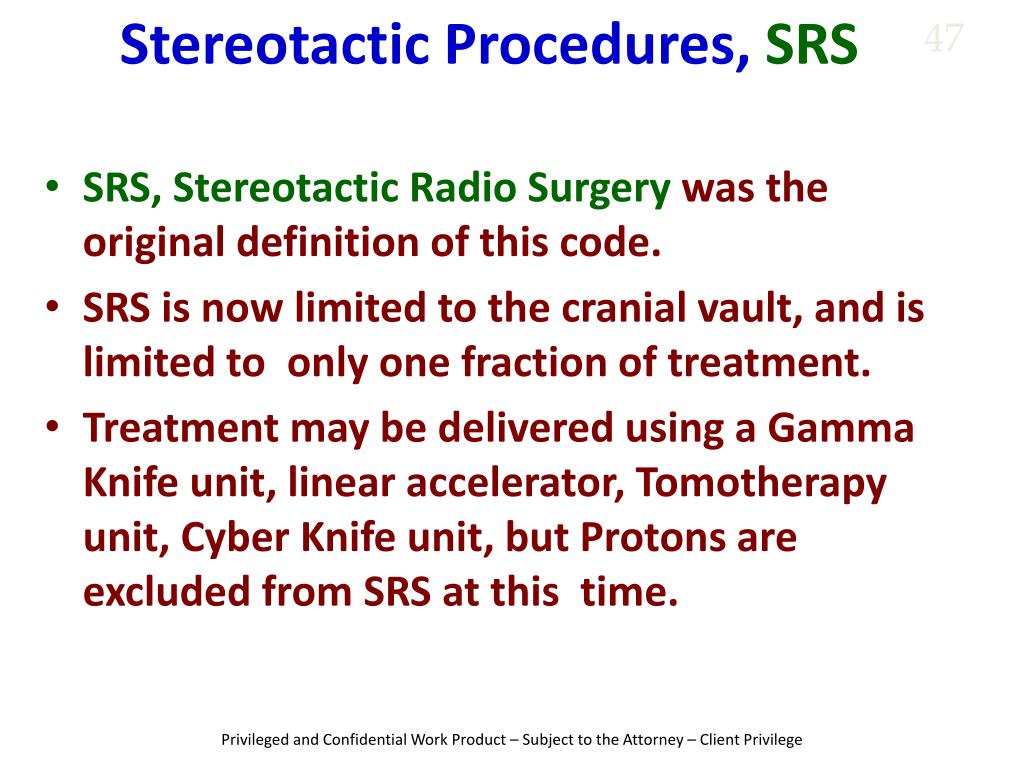 While both SRS and fractionated radiotherapy use ionizing radiation to treat brain tumors, their delivery methods, precision, and clinical applications differ significantly. The following table contrasts the two approaches:
Leave a Comment
Comments are moderated before appearing. The data you submit is processed according to the Privacy Policy of Findatopdoc.